
Health is a crown on the heads of the healthy that only the sick can see.


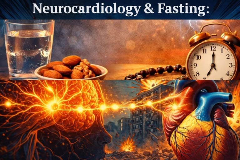
🧠🫀 Neurocardiology & Fasting : Unlocking the Heart–Brain Connection
Discover how fasting regulates the heart through the heart–brain axis. Learn how the autonomic nervous system and vagus nerve control cardiac function.
FASTINGNEUROCARDIOLOGY
Dr Hassan Al Warraqi
3/1/202610 min read


🧠🫀 Neurocardiology & Fasting : Unlocking the Heart–Brain Connection
Discover how fasting regulates the heart through the heart–brain axis. Learn how the autonomic nervous system and vagus nerve control cardiac function.
Your heart does not just beat to the drum of the brain.
It has its own nervous system — a sophisticated local network of neurons embedded within the heart tissue itself, capable of regulating rhythm and contraction independently of any signal from above.
This remarkable structure, known as the Intrinsic Cardiac Nervous System (ICNS), is the focus of one of medicine's most exciting emerging fields: neurocardiology.
Understanding the ICNS — and the broader heart-brain axis it belongs to — is transforming how we approach arrhythmias, stress-related heart disease, and the role of lifestyle interventions like fasting in cardiovascular health.
Key Insight: The heart is not just a pump.
It has local intelligence — and fasting may be one of the most accessible ways to protect and optimize it.
What Is the Neurocardiac Axis?
A Three-Level Control System
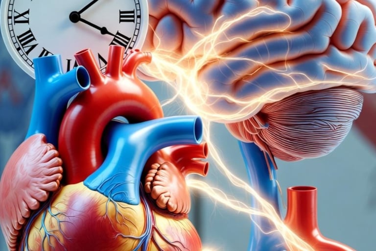
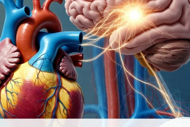
The neurocardiac axis is the bidirectional communication network linking the brain and heart.
It is organized into three hierarchical levels, each playing a distinct and complementary role in cardiac regulation
Level
Location
Primary Role
Level 1 (Central)
Medulla, Spinal Cord, Insular Cortex, Amygdala
High-level integration; emotional and stress responses; modulated by fasting via vagal afferents.
Level 2 (Peripheral)
Intrathoracic Ganglia (Stellate, Middle Cervical)
Relay station for sympathetic and sensory signals between brain and heart.
Level 3 (Intrinsic — ICNS)
Ganglionated Plexi in epicardial fat pads
Beat-to-beat modulation of heart rate, conduction, and contractility — independently of the brain.
What makes this system remarkable is its redundancy.
Even when Levels 1 and 2 are severed — as occurs in heart transplantation — Level 3 (the ICNS) continues to function, maintaining basic cardiac control autonomously.
This autonomy is both the heart's greatest strength and, under pathological conditions, a source of significant risk.
The 'Little Brain' of the Heart: Anatomy of the ICNS
The ICNS consists of thousands of neurons organized into Ganglionated Plexi (GP) — clusters of nerve cells embedded in the fat pads of the heart, primarily around the atria and near the pulmonary veins.
These are not passive relay points.
They contain three distinct functional neuron types:
Afferent (Sensory) Neurons: Act as the heart's internal sensors, monitoring mechanical stretch, pressure, and chemical changes in the myocardium.
Local Circuit Neurons (LCNs): The 'processors' of the ICNS. They integrate information from sensory inputs and from central nervous system signals, enabling intraganglionic processing.
Efferent (Motor) Neurons: The 'actuators' that translate processed signals into direct changes in heart rate (chronotropy), conduction velocity (dromotropy), and contractility (inotropy).
The ICNS is also neurochemically diverse — far more complex than once believed.
It contains both parasympathetic (cholinergic) and sympathetic (adrenergic) neurons, as well as a significant population of 'dual-phenotype' neurons expressing both acetylcholine and norepinephrine markers.
This complexity underlies the ICNS's ability to fine-tune cardiac function with extraordinary precision.
The Embryological Origin
Built Before the Brain
Cardiac innervation begins in the fifth week of human gestation.
Crucially, the parasympathetic system becomes functional before sympathetic differentiation is complete — a developmental sequence with profound implications.
Early in gestation, before either system is fully established, the heart relies on Intrinsic Cardiac Adrenergic (ICA) cells : a transient population that synthesizes catecholamines to sustain embryonic cardiac function.
A subset of these ICA cells persists into adulthood, forming a biological 'backup system' that becomes active during heart failure, ischemia, and post-transplant denervation.
Autonomic Imbalance : When the Heart-Brain Axis Breaks Down
Under ideal conditions, the sympathetic ('fight-or-flight') and parasympathetic ('rest-and-digest') arms of the autonomic nervous system operate in dynamic balance.
Chronic stress, overnutrition, poor sleep, and sedentary behavior tip this balance decisively toward sympathetic dominance.
The downstream consequences are well-documented and serious:
Reduced Heart Rate Variability (HRV) — the single most reliable non-invasive marker of autonomic health and neurocardiac resilience
Elevated resting heart rate and blood pressure
Endothelial dysfunction and accelerated atherosclerosis
Increased automaticity in the ICNS Ganglionated Plexi, particularly near the pulmonary veins
Higher incidence of atrial fibrillation, ventricular arrhythmias, and sudden cardiac death
The HPA axis plays a central role in this cascade.
Chronic psychological stress activates cortisol release, which further elevates catecholamine levels, reduces vagal tone, and creates a pro-arrhythmic neurocardiac environment.
This is not a metaphor — it is measurable physiology, with direct clinical consequences.
"The brain is the trigger; the heart is the target." — Bernard Lown, cardiologist. Fasting may help disarm the trigger.
How Fasting Influences the Heart-Brain Axis
Emerging research positions fasting as a meaningful non-pharmacological intervention targeting the neurocardiac axis at multiple levels simultaneously.
Its effects extend well beyond simple caloric reduction.
1. Autonomic Rebalancing : Restoring the Sympathovagal Balance
Multiple studies demonstrate that intermittent fasting (IF) and time-restricted eating (TRE) increase parasympathetic activity, measurable as improved HRV.
This shift is not merely a marker of wellbeing — it reflects a direct reduction in the excitability of the ICNS Ganglionated Plexi.
Specifically, fasting has been shown to lower circulating catecholamines (reducing the sympathetic burden on the ICNS), reduce resting heart rate (a direct indicator of enhanced vagal modulation), and improve HRV as a measurable sign of restored autonomic balance.
2. Metabolic Switching : Ketones as Cardiac Neuroprotectants
After approximately 12–16 hours of fasting, the liver begins producing ketone bodies, primarily beta-hydroxybutyrate (BHB).
This metabolic switch has profound effects on both cardiomyocytes and intracardiac neurons.
Ketones improve mitochondrial efficiency in cardiac muscle, reducing oxidative stress within the Ganglionated Plexi.
They also modulate ion channel activity — including KATP channels — which may stabilize electrical conduction and reduce ectopic firing.
Recent evidence further suggests BHB suppresses the NLRP3 inflammasome, reducing neuroinflammation within the ICNS itself.
3. BDNF Upregulation and Neuronal Resilience
Fasting activates cellular stress response pathways (AMPK, SIRT1, Nrf2) that enhance the production of Brain-Derived Neurotrophic Factor (BDNF).
Within the ICNS, BDNF supports neuronal survival, plasticity, and functional integrity — effects particularly relevant to age-related autonomic decline and post-transplant ICNS remodeling.
4. Circadian Alignment and Autonomic Rhythmicity
The Cardiac Autonomic Nervous System is deeply entrained to circadian rhythms.
Parasympathetic tone naturally peaks at night; sympathetic activity rises in the early morning — a pattern that aligns with peak periods for cardiac events.
Time-restricted eating (TRE) aligns food intake with natural light-dark cycles, reinforcing this circadian autonomic structure.
Late-night eating, by contrast, disrupts nocturnal parasympathetic dominance and elevates nocturnal sympathetic activity, increasing arrhythmia risk from ICNS hyperexcitability.
5. HPA Axis Modulation and Stress Resilience
Appropriately practiced fasting reduces cortisol reactivity to psychological stressors and dampens HPA axis overactivation.
This reduces the stress-induced sympathetic surges that trigger ectopic firing in the pulmonary vein Ganglionated Plexi — one of the primary mechanisms of atrial fibrillation initiation.
Fasting and ICNS Neurochemistry : Key Neurotransmitter Effects
Neurotransmitter
Fasting-Related Modulation
Cardiac Impact
Acetylcholine (ACh)
↑ Vagal tone enhances ACh release at sinoatrial node
Improved heart rate control; anti-arrhythmic effect
Norepinephrine (NE)
↓ Reduced sympathetic activity lowers NE spillover
Lower risk of tachyarrhythmias and ICNS hyperexcitability
Nitric Oxide (NO)
↑ eNOS activation via ketones and AMPK signaling
Vasodilation; improved coronary blood flow
Neuropeptide Y (NPY)
↓ Stress-induced NPY release reduced
Decreased sympathetic potentiation; less vagal suppression
VIP
May be upregulated with parasympathetic tone restoration
Supports post-vagal cardiac recovery; anti-fibrotic
Note: Individual autonomic responses to fasting vary significantly.
Patients with pre-existing conduction disease or autonomic neuropathy should consult a cardiologist before initiating any fasting protocol.
Clinical Implications : Fasting and Neurocardiac Disease
Atrial Fibrillation (AF)
Atrial fibrillation is the most common sustained cardiac arrhythmia.
Autonomic imbalance — particularly chronic sympathetic excess combined with episodic vagal surges — is a central driver, especially at the pulmonary vein Ganglionated Plexi.
Fasting addresses this through multiple converging mechanisms: reducing the sympathetic substrate that initiates ectopic firing, lowering systemic inflammation (CRP, IL-6) that sensitizes intracardiac neurons, improving metabolic substrate availability for stable electrical conduction, and reducing obesity and metabolic syndrome — independent major AF risk factors.
Observational studies of Ramadan fasting have reported meaningful reductions in AF recurrence and paroxysmal arrhythmia burden, providing early clinical support for this mechanism.
Sick Sinus Syndrome
An important and often overlooked clinical distinction : in some patients with Sick Sinus Syndrome, bradycardia is not caused by intrinsic sinus node failure but by excessive autonomic inhibition.
In these cases, autonomic blockade can restore a normal heart rate without any structural intervention.
Fasting's capacity to rebalance — not simply elevate — parasympathetic tone may help clarify and manage this subset of patients.
Neurogenic Stress Cardiomyopathy (Broken Heart Syndrome)
Takotsubo cardiomyopathy is triggered by a massive catecholamine surge from the brain, temporarily stunning the myocardium.
Lifestyle strategies like fasting that promote sustained sympathovagal balance may build resilience against such extreme neurocardiac events by reducing baseline sympathetic reactivity and stabilizing HPA axis responses to acute stress.
Heart Transplantation and Denervation
Heart transplantation severs the brain-heart axis at Levels 1 and 2.
The surviving ICNS undergoes remodeling, increasing its intrinsic neuronal excitability to compensate for the loss of extrinsic control.
Fasting-mimicking diets and ketosis may support this adaptation by enhancing neuronal survival via BDNF and reducing ischemia-reperfusion injury through upregulation of protective peptides like CGRP — expressed by the persistent ICA cells that serve as the heart's last autonomic backup.
Sudden Death in Epilepsy (SUDEP)
SUDEP illustrates the lethal consequences of a misfiring brain-heart axis.
Seizures trigger massive autonomic surges that can disrupt ICNS-regulated conduction, causing fatal arrhythmias.
The fasting connection is particularly relevant here: ketogenic diets, which share the metabolic state of prolonged fasting, have demonstrated reductions in seizure frequency — potentially also reducing the autonomic cardiac stress that accompanies seizures.
Fasting Protocols and Their Neurocardiac Effects
Fasting Protocol
Neurocardiac Benefit
12:12 Overnight Fast
Entry-level protocol; reduces nocturnal sympathetic surges; improves sleep-phase HRV
Intermittent Fasting (16:8)
Reduces catecholamine levels; enhances vagal tone; improves HRV and resting heart rate
Time-Restricted Eating (TRE)
Aligns feeding with circadian autonomic rhythms; reduces nocturnal arrhythmia risk
Prolonged Fasting (>24 hrs)
Triggers ketogenesis; reduces neuroinflammation in Ganglionated Plexi; BDNF upregulation
Caloric Restriction
Long-term reduction of sympathetic drive; lowers resting heart rate and blood pressure
Ramadan Fasting (observational)
Associated with reduced AF burden and paroxysmal arrhythmia recurrence
Integrative Management: Combining Fasting with Neurocardiac Care
Pharmacological Synergies
Fasting does not replace medication — it can complement it.
Beta-blockers combined with fasting may have additive effects on reducing sympathetic tone, though clinicians should monitor for excessive bradycardia.
SGLT2 inhibitors and fasting both promote ketosis and cardiovascular protection, with potential for enhanced metabolic benefit.
Aldosterone antagonists and fasting may jointly improve sympathovagal balance and reduce cardiac fibrosis.
Practical Protocol Guidance
Begin with a 12-hour overnight fast (e.g., 7pm to 7am) to reduce sympathetic nocturnal activity with minimal physiological stress
Progress gradually to 14–16 hour windows as tolerated, monitoring HRV as real-time feedback
Align eating windows with daylight hours to reinforce circadian autonomic entrainment
Break fasts with anti-inflammatory, magnesium-rich, omega-3-containing foods to support neuronal and cardiac membrane health
Combine fasting with aerobic exercise and mind-body practices (slow breathing, meditation, yoga) to amplify vagal tone benefits
Track resting heart rate, HRV, and inflammatory markers (hs-CRP) to individualize and assess response
Contraindications and Cautions
Fasting is not universally appropriate. Caution is essential in: advanced heart failure with cachexia, unstable arrhythmias or recent cardiac events, diabetes on insulin or sulfonylureas (hypoglycemia risk), eating disorders, and significant nutritional deficiencies.
Always consult a cardiologist before initiating extended fasting protocols.
Future Frontiers : Research at the Intersection of Fasting and Neurocardiology
ICNS-Specific Transcriptomics: Single-cell RNA sequencing of intracardiac neurons to map how fasting alters gene expression within Ganglionated Plexi and identify novel therapeutic targets.
Ketones as Direct Neuromodulators: Investigating beta-hydroxybutyrate's direct effects on GP excitability and neurotransmitter co-release dynamics.
Digital Phenotyping: Wearable ECG and continuous HRV monitors to enable real-time, personalized fasting protocols calibrated to individual autonomic responses.
iPSC Neurocardiac Co-Cultures: Patient-specific heart-neuron models to study fasting-induced metabolic states in hereditary arrhythmia syndromes like Brugada Syndrome.
Fasting-Mimicking Pharmacology: Developing compounds that replicate fasting's autonomic and anti-inflammatory benefits for patients who cannot safely fast.
Conclusion : Fasting as a Strategy for Heart-Brain Harmony
The heart-brain axis is one of biology's most sophisticated communication systems — and one of medicine's most underappreciated therapeutic targets.
The Intrinsic Cardiac Nervous System, the heart's own 'little brain,' is not a passive relay but an active, adaptive processing hub whose health is intimately tied to the metabolic and hormonal environment it operates in.
Fasting, practiced mindfully and individually, acts on this axis at multiple levels simultaneously: restoring the sympathovagal balance that modern life systematically disrupts, providing ketone-based neuroprotection to intracardiac neurons, aligning autonomic rhythms with circadian biology, and reducing the inflammatory milieu that drives ICNS hyperexcitability.
The message for clinicians, researchers, and patients is unambiguous: what you eat — and when — is not merely a metabolic question.
It is a neurocardiac one.
The heart has its own brain.
Fasting may be one of the most powerful, accessible ways to keep both in rhythm.
Frequently Asked Questions FAQS
Does fasting reduce the risk of atrial fibrillation?
Evidence from observational studies and mechanistic research suggests fasting can reduce AF burden by improving autonomic balance, lowering inflammation, reducing obesity, and stabilizing the excitability of pulmonary vein Ganglionated Plexi.
It is not a replacement for medical therapy but a potentially powerful adjunct.
What is the intrinsic cardiac nervous system (ICNS)?
The ICNS is a network of neurons embedded directly in the heart — primarily in the Ganglionated Plexi of the epicardial fat pads. Often called the heart's 'little brain,' it can regulate heart rate, conduction, and contractility independently of the central nervous system.
How does fasting improve heart rate variability?
Fasting reduces sympathetic overactivation and enhances parasympathetic (vagal) tone, shifting the autonomic nervous system toward a more balanced, resilient state.
HRV improves as a direct measurable consequence of this autonomic rebalancing.
Is fasting safe for people with heart disease?
Many patients with stable cardiovascular conditions can fast safely under medical supervision.
However, patients with advanced heart failure, unstable arrhythmias, insulin-dependent diabetes, or recent cardiac events should exercise significant caution and only fast under direct clinical guidance.
What role does the heart-brain axis play in stress-related heart disease?
Chronic psychological stress activates the HPA axis and sympathetic nervous system, flooding the heart with catecholamines.
This destabilizes the ICNS, reduces HRV, promotes endothelial dysfunction, and significantly elevates the risk of arrhythmias and neurogenic cardiomyopathy.
Fasting helps reduce this cascade by lowering cortisol reactivity and sympathetic baseline activity.
SEO Keywords: neurocardiology, heart-brain axis, intrinsic cardiac nervous system, ICNS, Ganglionated Plexi, fasting and heart health, intermittent fasting heart, autonomic nervous system, heart rate variability, atrial fibrillation fasting, vagal tone, sympathetic overactivity, cardiac arrhythmias, time-restricted eating heart, ketones cardiac health, circadian rhythm heart, fasting cardiovascular, ICNS fasting, neurocardiac junction, fasting HRV
© 2025 | Evidence-Based Neurocardiology Series | SEO-Optimized Clinical Content
========================================================================================================================================================================================
🧠🫀 Neurocardiology & Fasting : Unlocking the Heart–Brain Connection
https://www.h-k-e-m.com/-neurocardiology-and-fasting-unlocking-the-heart-brain-connection
Focus Keyword
neurocardiology heart-brain axis fasting
Secondary Keywords
intrinsic cardiac nervous system, ICNS fasting, intermittent fasting heart health, heart rate variability, autonomic nervous system heart, atrial fibrillation fasting, vagal tone, Ganglionated Plexi, cardiac arrhythmia lifestyle, fasting and heart health
Meta Description
Explore how neurocardiology, the heart-brain axis, and the intrinsic cardiac nervous system (ICNS) regulate cardiac health — and how fasting restores autonomic balance, reduces arrhythmia risk, and protects the heart's 'little brain'.
URL Slug
neurocardiology-heart-brain-axis-fasting
========================================================================================================================================================================================

Get in touch
Address
Cairo Al Rehab
Contacts
+20 109 405 2056
hassanalwarraqi@h-k-e-m.com